
Sonog-raphy shows the segmental type. The red curve represents the.
Other Malignancies In Thyroid Gland And Cervical Lymph Nodes Radiology Key
This study was designed to investigate the sonographic features of PTL.

. The left lobe of the thyroid measures 47cms craniocaudad by 17cms A-P by 15cms transverse which is normal in size. Hypervascularity is a typical finding in people with underlying autoimmune thyroiditis hashimotos or graves disease. Lymphoma is a disease in which lymphocytes ie.
Objective Primary thyroid lymphoma PTL is an uncommon thyroid malignancy. Thyroid lymphoma is a very rare disease that accounts for 1 to 2 of all thyroid cancers and 1 to 2 of all lymphomas outside the lymph nodes. A heterogeneous thyroid gland is seen in this 62 year old female patient.
Microcalcifications were found in 38 of cancerous nodules and only in 5 of benign non-cancerous nodules. Primary thyroid lymphoma PTL is a rare thyroid malignancy. This review article focuses only on primary.
This article is concerned with primary testicular lymphoma. Squamous cell carcinoma or lymphoma will seldom show a hilar line thought to be due to interruption of lymphatic flow by tumor invasion. Wang et alSpecific Sonographic Features of Primary Thyroid Lymphoma 320 J Ultrasound Med 2015.
Thyroid is a gland that serves several functions that affect the well being of the body. Primary Thyroid Lymphoma. Patients presenting with a palpable thyroid nodule is a common clinical dilemma.
479 cm 473 cm 395 cm 301 cm. Lymphoma is a cancer that develops in the lymphatic system the tissues and organs that produce store and carry white blood cells. Testicular lymphoma is an uncommon testicular malignancy.
B The color Doppler ultrasound shows reduction of twisted blood flow signals in the primary thyroid lymphoma after three cycles of rituximab plus bendamustine. The role of ultrasound in the assessment of cervical lymphadenopathy is well established. The incidence of solitary thyroid nodule is approximately 32 of the population in the UK and 42 in the USA 14They are four times more common in women than in men and the prevalence increases with age 14The risk of malignancy in a euthyroid patient with a.
Thyroid cancer was confirmed by thyroid color doppler ultrasound and puncture biopsy. Ad Learn more about the signs that may reveal you have an Issue that need attention. In thyroid lymphoma the lymphocytes of the thyroid turn into cancer cells.
Lymphoma of the Thyroid. Undefined 41 years experience. The patient is a 62 year female who had a biopsy proven lymphoma of the thyroid.
Primary thyroid lymphoma diffuse large B-cell type in a 66-year-old man presenting with rapidly growing neck swelling. This is called primary thyroid lymphoma to. The risk of cancer increased with the size of.
The 3 mm nodule is likely of. The present study describes the case of a patient who was confirmed to have PTL by intraoperative pathological. 11 Rago T Vitti P Chiovato L et al.
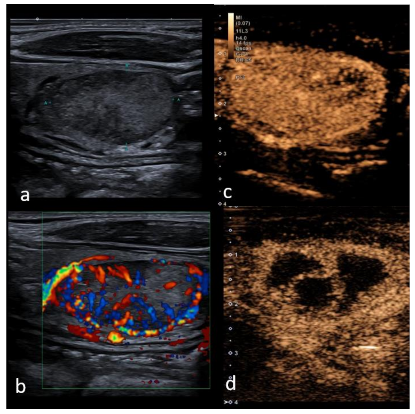
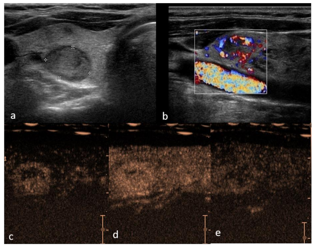
On spectral display a low resistance flow with a high peak systolic velocity is obtained. Thyroid cancer presenting as a hot thyroid nodule. Figure 4 Receiver operating characteristic curves of the combined contrast-enhanced ultrasound CEUS indicators in distinguishing primary thyroid lymphoma and nodular Hashimotos thyroiditis.
Lymphoma usually occurs within lymph nodes but in rare cases it arises from lymphocytes that are present within the thyroid gland. Methods Twenty-seven pathologically confirmed PTLs were. Thyroid lymphoma can classify as either primary or secondary thyroid lymphoma.
A The gray-scale ultrasonography shows no significant decrease of the tumor size transverse diameters. Primary thyroid lymphoma affects the thyroid gland first followed by spread to the lymph nodes and other organs later. Role of conventional ultrasonography and color flow-doppler sonography in predicting malignancy in cold thyroid nodules.
Report of a case and review of the literature. A Gray scale ultrasound transverse scan showing normal thyroid anatomy b Arterial vascularization of the thyroid gland. Primary thyroid lymphoma PTL is a rare thyroid malignancy.
On average 1 case of thyroid cancer was found for every 111 ultrasound exams performed. The black curve represents the combination of three ratios for peak intensity area under the timeintensity curve and time to peak. Primary manifestation of subclinical systemic disease.
Lymphoma can involve the testes in three ways. The purpose of this study was to determine the specific sonographic features of primary thyroid lymphoma and its color Doppler pattern compared to nodular goiter. Primary site of extranodal disease primary testicular lymphoma secondary involvement of systemic disease.
The hilar line is more prominent in older patients. Lymphoma can involve in multiple lymph nodes and extranodal organs including the gastrointestinal tract thyroid gland breast and bones. In accordance with the suggested.
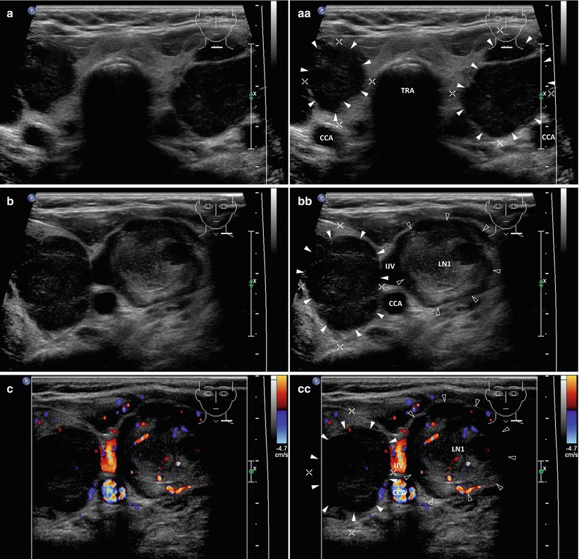
Color Doppler imaging shows a cen-. Most patients are older with an average age in the late 60s. Clinical diagnosis of PTL may not be easily established based on imaging studies as the imaging features of PTL are similar to those of lymphocytic thyroiditis and primary thyroid cancer.
It is particularly sensitive compared to clinical examination 968 and 733 respectively in patients with previous head and neck cancer with post-radiation neck fibrosis. Malignant lymph nodes in the neck whether they are metastatic from the thyroid or elsewhere ie. Secondary thyroid lymphoma affects lymph nodes and other organs first followed by subsequent spread to the thyroid.
Clinical diagnosis of PTL may not be easily established based on imaging studies as the imaging features of PTL are similar to those of lymphocytic thyroiditis and primary thyroid. Thyroid nodules were found in 97 of patients with thyroid cancer and in 56 of without thyroid cancer. Despite the rarity of PTL it is important to recognize PTL promptly because its management differs from that of all the other thyroid neoplasms.
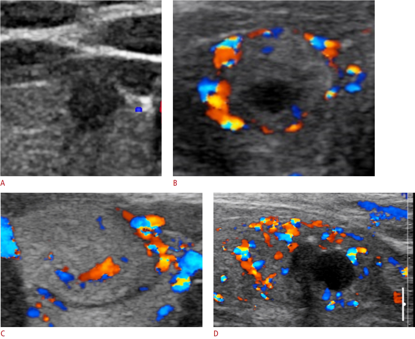
Blood cells that fight infection turn into cancer cells. On color Doppler the inferior thyroid artery arrow is seen c Blood flow pattern in normal thyroid gland. The sonographic findings for 13 surgically proven primary thyroid lymphomas were analyzed and compared to those for 27 nodular goiters.

Application Of Color Doppler Ultrasound To Evaluate Chemotherapeutic Effect On Primary Thyroid Lymphoma
Usg Ultrasonography Ultrasonography 2288 5919 2288 5943 Korean Society Of Ultrasound In Medicine 10 14366 Usg 20072 Usg 20072 Review Article Clinical Applications Of Doppler Ultrasonography For Thyroid Disease Consensus Statement By The

Primary Thyroid Lymphoma Has Different Sonographic And Color Doppler Features Compared To Nodular Goiter Wang 2015 Journal Of Ultrasound In Medicine Wiley Online Library
Cancers Free Full Text Performance Of Contrast Enhanced Ultrasound In Thyroid Nodules Review Of Current State And Future Perspectives Html

Thyroid Lymphoma Nam 2012 Journal Of Ultrasound In Medicine Wiley Online Library
Cancers Free Full Text Performance Of Contrast Enhanced Ultrasound In Thyroid Nodules Review Of Current State And Future Perspectives Html
Primary Thyroid Lymphoma Has Different Sonographic And Color Doppler Features Compared To Nodular Goiter Wang 2015 Journal Of Ultrasound In Medicine Wiley Online Library

Application Of Color Doppler Ultrasound To Evaluate Chemotherapeutic Effect On Primary Thyroid Lymphoma

Primary Thyroid Lymphoma Has Different Sonographic And Color Doppler Features Compared To Nodular Goiter Wang 2015 Journal Of Ultrasound In Medicine Wiley Online Library